


By: Prof. Dr. Seyed Saeid Zamanieh Shahri, MD and Prof. Dr. Sonia Sayyedalhosseini, MD
Bladder Cancer:
Cancer is the abnormal growth of malignant cells in the body. Bladder tumors usually develop in the bladder wall. Bladder is a part of the reproductive system where urine is collected and then discharged. Bladder cancer is usually curable if it is in its early stages, not to mention that many cancers are curable if detected early.
Bladder Anatomy and Physiology:
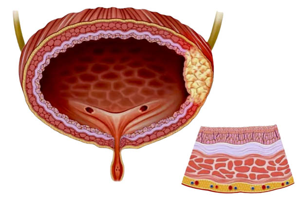
The bladder is a muscular bag in the pelvis that is located just above and behind the pubic bone. The appearance of the bladder varies depending on the amount of urine stored in it, and when it is full, it is oval in shape, and when it is empty, it is the size and shape of a pear. The bladder is lined by layers of muscle tissue that are elastic to hold urine. The inner tissue of the bladder is folded into the bladder and gives it an elastic and flexible property. When the bladder empties, it contracts again and returns to its normal state, and the wall of the bladder muscle becomes thicker and the entire bladder hardens. The external structure of the bladder includes:
Apex, which is connected to the umbilicus by the median ligament (remains of the urethra).
The body, which is the main part of the bladder and is located in its upper part.
The base or fundus is a triangular structure located at the back or end of the bladder.
The neck is connected to the urethra.
Bladder muscles:
Bladder muscles play a key role in storing and emptying urine. The bladder wall contains a specialized smooth muscle known as the detrusor muscle, which maintains the integrity of the bladder structure during stretching and causes urination during bladder contraction. There are also two muscular sphincters in the urethra: internal urethral sphincter, external urethral sphincter
The female urethral sphincter is a functional sphincter and has no muscles. The internal urethral sphincter in men consists of circular smooth fibers, which are under the control of voluntary nerves and prevent the sperm from reabsorbing during ejaculation.
The external urethral sphincter has the same structure in men and women. This sphincter is a skeletal muscle and is under the control of involuntary nerves. However, the mechanism of the external sphincter in men is more complex.
In terms of structure and function, the bladder is an organ of the urinary system that plays two main and important roles in the body:
• Temporary storage of urine
Bladder is a hollow organ with flexible walls, which in adults allows the bladder to store 400 to 600 ml of urine temporarily until emptying. Depending on the amount of urine accumulation, the size of the bladder can increase. Normally, the bladder holds 400 to 600 milliliters of urine, but when it is about a quarter full, a person feels the need to urinate.
• Help with urination
The structure and function of the bladder is such that it can play an effective role in the excretion of urine. Urine is made in the kidneys and transported to the bladder through two tubes called the ureters. The bladder stores urine and controls its excretion. When the bladder is full, it releases urine through the urethra. In women, this tube ends between the clitoris and the vagina. During urination, the bladder muscles contract and two sphincters open to allow urine to leave the body. The internal sphincter in the bladder is a muscular valve that prevents leakage of urine. The trigone in the bladder is a triangular base that prevents stretching of the urethra or backflow of urine into the urethra.
Types of bladder cancer: There are three types of bladder cancer.
– Transitional Cell Bladder Cancer: Transitional cell cancer is the most common type of bladder cancer. It begins in transitional cells in the inner layer of the bladder. Transitional cells are cells that change shape when the tissue is stretched without being damaged.
– Squamous Cell Bladder Cancer:
Squamous cell cancer is a rare cancer in the United States. This disease begins when thin, flat squamous cells form in the bladder after a long-term infection or bladder irritation.
– Adenocarcinoma Bladder Cancer: Adenocarcinoma is also a rare cancer in the United States. This disease begins when glandular cells form in the bladder after long-term irritation and inflammation of the bladder. Glandular cells form mucus-secreting glands in the body.
The beginning and spread of bladder cancer:
The bladder wall has several layers. Each layer is made up of different cells. Most bladder cancers begin within the inner layer of the bladder, called the urinary or transitional epithelium. As cancer grows into or through other layers of the bladder wall, it is more advanced, and more difficult to treat. Over time, cancer may grow outside the bladder and into nearby structures. It may spread to nearby lymph nodes or other parts of the body. (When bladder cancer spreads, it tends to go to distant lymph nodes, bones, lungs, or liver.)
Aggressive and non-aggressive Bladder Cancer:
Bladder cancers are often described based on how far they have spread into the bladder wall:
Non-invasive cancers are only in the inner layer of cells (transitional epithelium). They have not grown into deeper layers.
Invasive cancers have spread to deeper layers of the bladder wall. These cancers are more likely to spread and are more difficult to treat.
Bladder cancer can also be described as superficial or non-muscle invasive. These terms include both non-invasive tumors and invasive tumors that have not grown into the main muscle layer of the bladder.
Risk factors:
Smoking increases the risk of bladder cancer. The following factors also increase the risk of developing bladder cancer:
Exposure to cancer-causing chemicals, chronic bladder infections, low fluid intake, being male, being white, being older, since most bladder cancers occur in people over 55, having a high-fat diet, having a family history of bladder cancer, previous treatment with certain chemotherapy drugs, having previous radiation therapy to treat cancer in the pelvic area, arsenic in drinking water, certain medications, exposure to workplace pollutants and etc.
Race and ethnicity, age, chronic bladder irritation and infection, personal history of bladder or other urinary cancers, birth defects, genetics, and family history.
Causes of bladder cancer:
This cancer starts when changes (mutations) occur in the DNA of bladder cells. A cell’s DNA contains instructions that tell the cell what to do. But these changes tell the cell that when the time comes for healthy cells to die, they multiply quickly and continue to live. Abnormal cells gradually form a tumor that can attack and destroy normal body tissue. Over time, abnormal cells can spread and spread throughout the body (bladder cancer metastasis).
Signs and symptoms:
Visible symptoms of bladder cancer and its symptoms may include the following:
Blood in the urine (haematuria), which may cause the urine to appear bright red or the color of Coca-Cola, however sometimes the urine looks normal and the presence of blood is only detected in a laboratory test.
Frequent urination, Painful urination, Back ache
Diagnosis:
Tests and methods used to diagnose this cancer may include the following:
• Using a scope to examine the inside of the bladder (cystoscopy). To perform a cystoscopy, doctor inserts a small, narrow tube (cystoscope) through the urethra. A cystoscope has a lens that allows doctor to see inside urethra and bladder, to check these structures for signs of disease. A cystoscopy can be done in a doctor’s office or hospital.
• Taking tissue samples for testing (biopsy). During a cystoscopy, doctor may pass a special instrument through a scope into the bladder to collect a sample of cells for testing (sampling). This method is sometimes called bladder tumor sampling through the urethra. It can also be used to treat bladder cancer.
• Urine sample examination (urine cytology). A sample of urine is analyzed under a microscope to check for cancer cells in a procedure called urine cytology.
• Imaging tests, such as a CT urogram or retrograde pyelogram, allow doctor to examine the structures of urinary tract. During a CT urogram, the contrast material injected into a vein in arm eventually flows into kidneys, ureters, and bladder. X-ray images taken during the test provide a detailed view of urinary tract and help doctor identify areas that may be cancerous. A retrograde pyelogram is an X-ray examination used to closely examine the upper urinary tract. During this test, doctor will pass a thin tube (catheter) through your urethra and bladder to inject contrast material into ureter. While x-ray images are taken, dye is injected into the kidneys.
• After bladder cancer is confirmed, the doctor may recommend other tests to determine if the cancer has spread to the lymph nodes or other areas of the body.
These tests may include the following:
CT scan, magnetic resonance imaging or MRI, positron emission tomography or PET scan, bone scan, chest X-ray.
Doctor uses the results of these methods to determine the stage of the cancer. Bladder cancer stages are indicated by Roman numerals from 0 to 4 (zero to four). The lowest stages indicate cancer that is confined to the inner layers of the bladder and has not yet grown enough to affect the muscle wall. The highest stage, stage IV, indicates cancer that has spread to lymph nodes or distant organs in the body.
Grading of bladder cancer:
Bladder cancers are further classified based on how the cancer cells appear when viewed through a microscope. This is known as grading, and your doctor may describe the cancer as low grade or high grade:
Low-grade Bladder Cancer: This type of cancer has cells that are closer to normal cells in appearance and organization (well-differentiated). A low-grade tumor usually grows more slowly and is less likely to invade the muscle wall of the bladder than a high-grade tumor.
High-grade Bladder Cancer:
This type of cancer has cells that have an abnormal appearance and do not resemble normal-looking tissue (not much different). A high-grade tumor tends to grow faster than a low-grade tumor and may spread to the muscular wall of the bladder and other tissues and organs.