


By: Prof. Dr. Seyed Saeid Zamanieh Shahri, MD and Prof. Dr. Sonia Sayyedalhosseini, MD
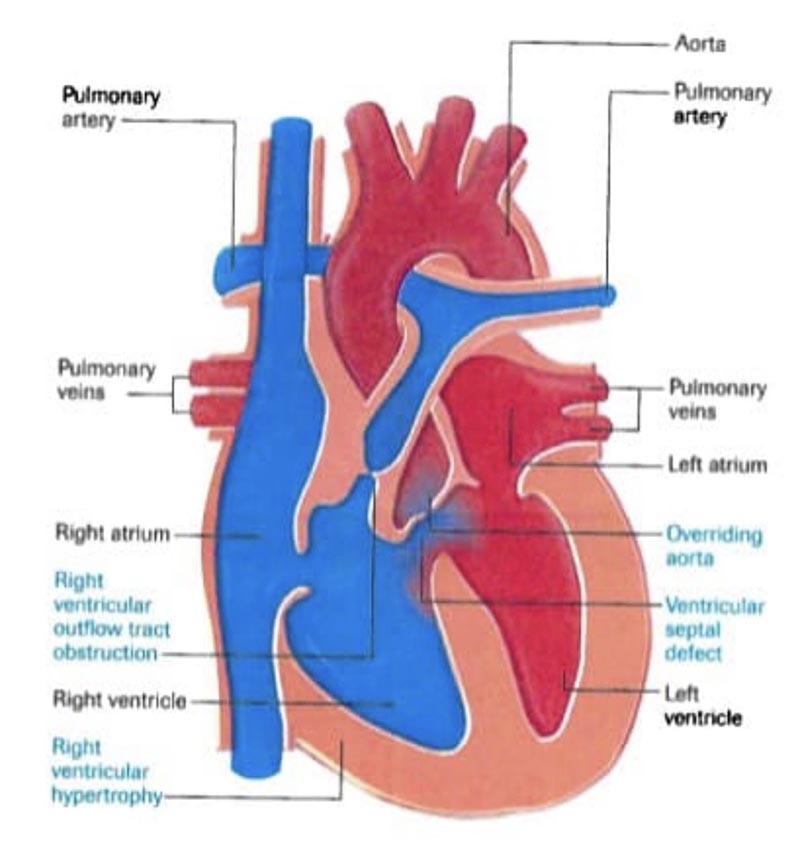
Tetralogy of the Fallot is a congenital defect that affects the normal flow of blood from the heart to other parts of the body. It occurs when the baby’s heart is not properly formed and the baby is not growing enough in the uterus. Tetralogy of Fallot is a collection of four defects in the heart and arteries. These defects include: ventricular wall defects, pulmonary valve stenosis, right ventricular hypertrophy, and abnormal location of the aorta.
The presence of these defects reduces the oxygen flow from the heart to the body. Because of this, babies with this condition have bluish skin. Of course, not all babies may have blue skin, and only parts of the body, turn blue due to cyanosis when crying or feeding.
Anatomy of the heart:
The human heart has two main cavities at the top and two cavities at the bottom. Each of these cavities has its own function and is separated by walls. The right ventricle, located in the lower part of the heart, sends oxygen-free blood to the lungs, and the left ventricle receives oxygenated blood from the lungs. This blood is then carried by the aorta throughout the body. Any defect in this structure disrupts heart function and causes disease. Tetralogy of Fallot is one of the rare congenital diseases caused by the four defects mentioned above in the heart.
Pathophysiology of tetralogy of Fallot:
As mentioned, the normal heart has four chambers. The two upper chambers of the heart are called the atria and the lower chambers are called the left and right ventricles. The ventricular wall separates the ventricles and prevents the combination of oxygenated and deoxygenated blood. In a normal heart, blood travels from the right ventricle through the pulmonary artery to the lungs, where it receives oxygen. Oxygenated blood then returns to the left ventricle and is sent throughout the body through the aorta (the body’s main artery). But as mentioned, the heart with tetralogy of Fallot has four defects that can be explained as follows:
Interventricular wall defect:
The presence of a defect in ventricular wall causes a mixture of oxygenated blood (blood in the left ventricle) and deoxygenated blood (blood in the right ventricle).
Pulmonary valve stenosis:
This defect prevents the pulmonary valve from opening fully. In this case, blood flows from the right ventricle to the pulmonary artery which is restricted. Therefore, the heart must be more active to be able to pump blood to the lungs with more pressure.
Right ventricular hypertrophy:
In this condition, the right ventricular muscle becomes larger and thicker than the left side of the heart, due to excessive blood flow.
Abnormal placement of the aorta: In a normal heart, the aorta is attached to the left ventricle, allowing oxygen-rich blood to flow throughout the body. In tetralogy of Fallot, the aorta is located between the left and right ventricles. This causes hypoxic blood to enter the aorta instead of the pulmonary artery.
Causes:
The exact cause of tetralogy of Fallot is unknown. However, some studies suggest that the disorder may be due to the interaction of several genetic and environmental factors with each other or some factors alone.
Therefore, researchers believe that factors may affect the developing fetus’s genes and cause these congenital defects. But the exact nature of these factors is unclear. Extensive research into rare diseases, viral diseases, alcohol use, diabetes, malnutrition, and pregnancy over the age of 40 increases a child’s chances of developing tetralogy of heart failure.
pproximately 25% of infants with the disease have other birth defects that are not related to heart function or structure.
Symptoms of tetralogy of Fallot in infancy and childhood:
Symptoms of tetralogy of Fallot occurs more in infancy and childhood.
Abnormal heart sounds or cyanosis in the first few weeks of life are much more common than other symptoms. The degree of cyanosis or bluish skin depends on the amount of blood flow to the lungs. Also, due to low oxygen levels, the baby may have difficulty breathing.
In addition, other symptoms that may occur in infants is anemia, abnormal increases in the number of red blood cells in children related to the hypoxia, and the passage of blood clots through the bloodstream are some of these symptoms. These clots may stop blood flow to the brain and cause strokes.
Symptoms of tetralogy of Fallot in adults:
People with tetralogy of Fallot are usually treated in childhood. So many adults with the disease are usually asymptomatic. But they may still have to deal with the problems. Adults with tetralogy of Fallot may have symptoms such as irregular heart rhythm and blockage of blood on the right side of the heart. They also have limited ability to exercise and do strenuous activity.
Complications: After surgery, the lung valve may still be blocked or leaked. In this case, the child’s physical activity is limited, especially in competitive sports. Children with tetralogy of Fallot may also have decreased heart function or may have heart rate disorders. People with this disease usually need to see a cardiologist regularly. This trend should continue into adulthood. Some of these patients’ problems may get worse in the long run and increase the need for more medication and surgery or even catheterization (insertion of a catheter or valve into a heart vessel).
Diagnostic methods: Tetralogy of Fallot can be diagnosed during pregnancy or immediately after birth. Screening tests are done before birth to check for birth defects and other diseases. Symptoms of the disease may also be seen on ultrasound.
In this case, the doctor will prescribe a Fallot test for the mother to make sure it exists. A fetal echocardiogram is an ultrasound of the fetal heart that can show problems with the structure and function of the fetal heart. Tetralogy of Fallot is diagnosed after the baby is born, usually with blue skin when feeding or crying. Hearing loud and abnormal sounds from the baby’s heart by stethoscope at birth is another way to diagnose heart with Fallot tetralogy. An echocardiogram at this time can also be helpful in diagnosing the disease.


