


By: Prof. Dr. Seyed Saeid Zamanieh Shahri, MD and Prof. Dr. Sonia Sayyedalhosseini, MD
Atrial Septal Defect (ASD) is a hole in the wall between the two upper chambers of the heart (atria). This defect can be present at birth and is actually a congenital disease.
Minor defects may never cause a problem or symptom and may even be diagnosed accidentally. It may resolve spontaneously after childbirth or during childhood.
Atrial septal defect, if large and advanced, can damage the heart and lungs. People with congenital heart disease who have had symptoms for years have a poor quality of life. Because this disease affects the heart and pulmonary arteries. Therefore, surgical treatment is needed to improve the living conditions of these people.
Symptoms of atrial septal defect:
Many babies born with atrial septal defects do not have the associated signs and symptoms. In adults, the signs or symptoms may begin around the age of 30, but in some cases the signs and symptoms may not appear until decades later.
Signs and symptoms of atrial septal defect may include:
• Shortness of breath, especially during exercise
• Early fatigue
• Swelling of the toes, abdomen or legs
• High heart rate or palpitations
• heart attack
• Heart murmur
* Causes of atrial septal defect:
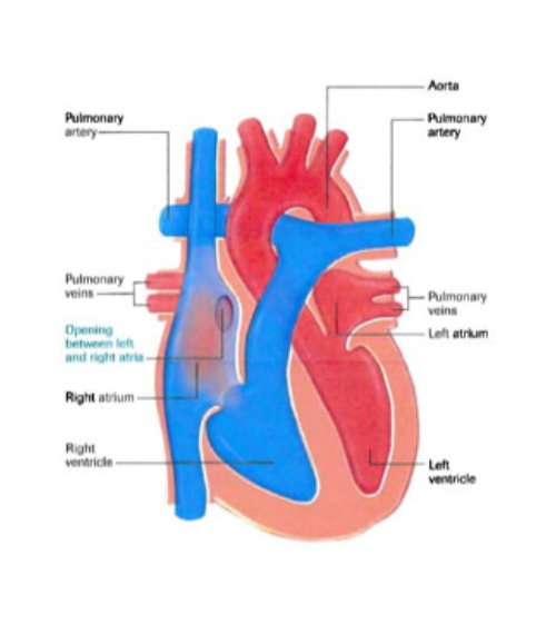
The heart is divided into four hollow cavities, two on the right and two on the left. To pump blood throughout the body, the heart uses its left and right to perform various functions.
The right side of the heart carries blood to the lungs through the pulmonary arteries. In the lungs, the blood absorbs oxygen and then returns to the left side of the heart through the pulmonary arteries. On the left side of the heart, blood is sent throughout the body through the aorta (the body’s largest artery).
Doctors believe that the cause of this defect is related to the developmental period of the heart during fetal period, But they usually do not give a specific reason for it. However, genetics and environmental factors may play a role in the development of atrial wall defects.
How does a heart with an atrial wall defect work? As mentioned, the defect of the atrial wall is the creation of holes between the walls of the atria. Thus, during this defect, the oxygen-rich blood of the left atrium mixes with the deoxygenated blood by flowing to the right atrium. In this case, the mixed blood is pumped back to the pulmonary arteries.
If the atrial wall defect is severe, the volume of blood flowing through the pulmonary arteries increases, increasing the activity of the heart. If left untreated, it can overwhelm the right side of the heart over time and the right ventricle becomes weak due to overwork. Continuing this process also increases blood flow to the pulmonary arteries, causing pulmonary hypertension..
Types of atrial septal defects:
There are three common types of atrial septal defect (ASD) and one uncommon type:
Ostium Secundum: Located in the center of the atrial septum, which is the most common type of ASD.
Ostium Premium: This defect is located near the lower part of the atrial wall, may be associated with defects in the mitral and tricuspid valve (the second most common type).
Sinus Venusus: Located near the top of the septum between the atria and is often associated with an abnormal connection of the right pulmonary vein (s) to the right atrium instead of the left atrium.
Coronary Sinus: In this rare defect, part of the wall between the coronary sinus (which is part of the heart’s vascular system) and the left atrium is lost.
In atrial septal defect, there is usually more pressure in the left atrium than in the right atrium, so blood flows from the left to the right (shunt). Surgical medical advice for ASD closure depends primarily on the characteristics of the defect (size, location), the symptoms or limitations of the defect, and the preferences of the patient and physician.
To be Continued…


