


By: Prof. Dr. Seyed Saeid Zamanieh Shahri, MD and Prof. Dr. Sonia Sayyedalhosseini, MD
The size and shape of each cell depends on its function. The muscle cell is long so that it can move the skeleton.
The nerve cell is also long so that it can transmit nerve signals quickly from one point to another in the body. The length of a muscle cell may reach several centimeters and the length of a nerve cell up to a meter. The red blood cell is small and its diameter is only 7.5 to 8 microns, and for this reason it can pass through the narrowest vessels of the body.
The smallest cell is mycoplasma, which is a prokaryote without a wall and is a common contaminant of cell culture media. The diameter of mycoplasma is only 0.3 microns. Mycoplasma is saprophytic or lives parasitically in higher organisms.
Some cells have completely different shapes, for example, a giant cell is a large cell with a variable shape, large cytoplasm and multiple nuclei, like an osteoclast cell or the bone-eating cell that is responsible for bone destruction, it is formed from the close cooperation of several individual cells, with a uniform cytoplasmic mass in which there are several nuclei. Or like a syncytiotrophoblast cell in the human placenta.
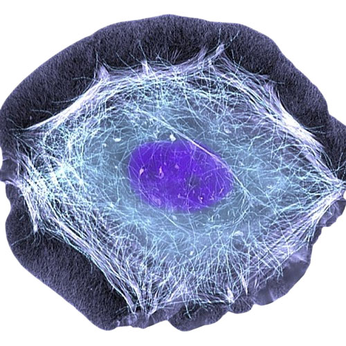
The limiting factor in cell size is the surface area to volume ratio, that is, the cell surface area must be large enough to be able to absorb sufficient nutrients from the environment and eliminate waste products. In this way, the cell size is larger than a certain limit and does not become smaller than a certain limit. A larger cell has a larger surface area, but its surface area to volume ratio is smaller compared to a smaller cell of the same shape.
Therefore, it can be concluded that the surface area to volume ratio is smaller in a larger cell and vice versa. Obviously, the larger the surface area to volume ratio of the cell, the easier it is to meet the needs of the cell and vice versa.
Another limiting factor in cell size is the ratio of the volume of the nucleus to the volume of the cell. In organs with intense metabolic activity, cells are smaller so that the time for diffusion of substances is reduced. Examples of these small and active cells are embryonic cells.
Life and death of cells:
The normal lifespan of cells in a healthy human body varies from a few days to eighty years. In each period of life, the cell is initially in an embryonic state, meaning that the cytoplasm is small and absent and it has a large nucleus.
Such an embryonic cell, which is multipotential, is called an undifferentiated cell or an immature cell, such as a mesenchymal cell. After the embryonic stage, the cell reaches the maturation stage, meaning that the cell growth is complete and it acquires a specific shape and function. In this case, it is called a differentiated cell or an adult cell. Sometimes an undifferentiated cell may become regular and organized through a process of regulation or modulation and later create a specific tissue or lineage, that is, it becomes differentiated. Sometimes a differentiated cell returns to its pre-mature state, which is called dedifferentiation, such as when cartilage turns into mesenchyme. A progenitor cell has an extraordinary ability to proliferate and creates a large number of final cells, but its self-renewal power is usually low. A stem cell is a dormant and inactive progenitor cell that has both a high ability to proliferate and, to a greater extent, the power to self-renew. In some situations, such as hematopoiesis, the cell is able to transform into different types of differentiated cells, and for this reason it is called a multipotent or pluripotent cell. In contrast, a unipotent or pluripotent cell is only able to create one type of cell, such as sperm. Differentiation, proliferation, and proliferation are three different terms that will be explained.
One of the interesting phenomena in the precise and correct performance of cell functions is the induction phenomenon, which is a phenomenon under the control and influence of which cells begin some of their functions. This phenomenon causes cells in the human embryo to differentiate and create a special tissue. The induction phenomenon affects cells that meet the conditions for induction not only in the embryonic period but also in postnatal life. That is, in cases of necessity, it forces some cells to begin differentiation and create new differentiated cells, and these new cells also act according to the previous commitment that has been induced to them. In this way, if a bone-forming cell is transplanted to a muscle, the cell does not build muscle, but builds bone according to its principle and commitment. Cell death may be the result of a physiological process that leads to a dead cell, that is, a cell dies after a normal life and gives its place to another cell. This type of cell death is called physiological death or programmed death or apoptosis, such as the death of cells in the outer layer of the epidermis in the skin.
Another type of cell death is pathological death, which leads to necrotic cells, that is, a cell dies as a result of a pathological condition, such as mechanical damage or a toxin. This type of cell death is called necrosis. Although the cytoplasm also undergoes changes in a dead cell, changes in the nucleus usually attract more attention. The changes in the cytoplasm and nucleus in apoptosis and necrosis have similar stages, which are:
* Cytolysis: The cytoplasm thins, the cell swells, and then bursts.
*Coagulation: The cytoplasm thickens and becomes a special, irreversible gelatinous substance
*Pycnosis: The nuclear chromatin becomes dense and crumpled, forming a dense, compact mass
*Karyorrhexis: The nucleus becomes fragmented and scattered.
*Karyolysis: The nucleus loses its ability to stain and disappears so completely that it appears to have dissolved. This stage is also called chromatolysis due to the loss of chromatin staining ability. So, there are two types of cell death. Physiological death. Pathological death.


