By: Prof. Dr. Seyed Saeid Zamanieh Shahri, MD and Prof. Dr. Sonia Sayyedalhosseini, MD
*Exocytosis: What remains in the digestive vesicle after digestion is called the residual body, which contains indigestible materials and is often excreted through the cell membrane by the phenomenon of exocytosis.
*Functions of lysosomes: Phagosome is a membrane-bound vesicle in the cells like macrophages, which contains xenobiotic materials. The contents of the phagosome can be foreign materials that have entered the cell from the outside, in which case it is called a heterophagosome, but if the contents of the phagosome are damaged or worn-out structures of the cell, it is called an autophagosome. Heterophagosome is formed as a result of xenobiotics and autophagosome as a result of autophagy (self-eating) of the cell.
If damage to cells causes rupture of lysosomes inside the cell, the released enzymes immediately begin to digest adjacent organic materials. If the cell injury is mild, only part of the cell is removed and the cell is subsequently repaired. If the cell injury is severe, the entire cell is digested, which is called autolysis. The removed cell is replaced by a new cell of the same type, usually by mitotic proliferation.
Sometimes body tissues degenerate and shrink, such as shrinking of the uterus after childbirth, or muscle atrophy due to immobility or shrinking of the breasts after breastfeeding.
It is interesting to know that lysosomes are responsible for a large part of tissue degradation, but the mechanism by which the lack of activity of a tissue increases the activity of lysosomes is not clear.
Lysosomes contain bactericidal enzymes and substances, so they can kill phagocytic bacteria before they cause cellular damage.
These enzymes and substances include: lysozyme, lysoferrin, and various acids. Lysozyme dissolves the bacterial cell membrane. Lysoferrin absorbs iron and other metals essential for bacterial growth, and an acid with a pH below five activates hydrolases and disables some of the bacterial metabolic systems. It is now easy to conclude that lysosomes can digest vesicles ingested by the cell, degrade tissues, cause autolysis of cells, and kill phagocytic bacteria.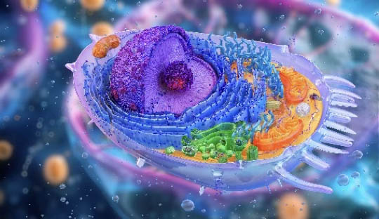
*Mitochondria: Mitochondria are found in all aerobic eukaryotic cells. The word mitochondria are derived from mito, meaning thread, and chondrion, meaning granule. The depressions in the inner membrane of mitochondria are called cristae.
Mitochondria are the powerhouse of the cell, or the energy-generating device of the cell, or the site of cellular respiration. Without mitochondria, the cell cannot extract the energy it needs from food, and as a result, virtually all cellular functions stop. Most of the energy obtained from respiratory oxidation is trapped in this organelle.
Mitochondria, which combine respiration with the production of the high-energy intermediate substance ATP, undergo a process called oxidative phosphorylation. After digestion and absorption, they convert carbohydrates, proteins, and fats in food into their building blocks.
Mitochondria in human cells are special organelles whose important function is to firstly possess the enzymes necessary to produce energy from glucose, fatty acids, and amino acids, and secondly, they use this energy to obtain the more important and energy-rich compound in the form of ATP (Adenosine Tri-Phosphate) from a precursor called ADP (Adenosine Di-Phosphate). End
The structures inside the cell:(1)
*Centrosome: In the vicinity of the nucleus, two rod-shaped structures are observed, each of which is called a centriole, and their set is called a centrosome, which are usually in pairs and are located in the center of the cell. Centrioles participate in the process of cell division. With an electron microscope, each centriole is seen as a cylindrical structure with one side open and the other closed. The wall of the centriole consists of 9 hollow 3-stranded microtubules that are located at equal distances from each other and in an amorphous matrix. Each group of thirty of the nine groups of microtubule structures is called a triplet. The motor appendages of unicellular organisms, such as cilia and flagella, also have a structure similar to the centriole and are studied as derivatives of the centriole. In fact, the centriole is composed of 9 groups of 3-stranded microtubules, each of which is called a triplet. Cilium and flagellum are the motor organs of the cell. A cilium is a tiny hair-like appendage that protrudes from the surface of the cell. The rhythmic movement of cilia, like the movement of a breeze in a wheat field, causes the cell to move or the movement of fluid or mucus. A flagellum is a long, mobile appendage that protrudes from the surface of the cell. Flagellin is the protein of the bacterial flagellum. The central core of a cilium or flagellum, which has 2 central microtubules and 9 paired peripheral microtubules, is called an axoneme. From each doublet, two protein arms of the dynein type separate towards the next doublet, which do not reach the next doublet, and each doublet is connected to the next doublet by horizontal filaments of the protein nexin. Dynein is an analogue of myosin and doublet is an analogue of actin in muscle. Here, like in muscle, a sliding mechanism causes the movement of the cilia or flagellum, which is similar to the sliding mechanism of actin and myosin in muscle.
* Cytoskeleton: The cytoskeleton is made of protein strands called microtubules, microfilaments, and centrioles.
*Microtubules: Microtubules or small tubes are a type of cytoplasmic organelle that are made of small, strong, flexible tubes in the shape of a pipe or rod called microtubules. These tubes are located in the cytoplasm as diffuse components and are made of the protein tubulin. In general, microtubules form the fundamental component of the cytoskeleton and play an important role, especially in unicellular organisms and flagellates. Microtubules strengthen various parts of the cytoplasm. The spindle apparatus during mitosis, the sperm tail coating, and the marginal filaments in a nucleated red blood cell are made of microtubules. The inner part of the microtubule is less dense and appears hollow. In addition to keeping parts of the cytoplasm strong and stable, microtubules also help to maintain the shape of the cell. A cross-section of a microtubule shows that the wall of each microtubule is made of 13 smaller, parallel tubes called protofilaments. Microtubules are made of two different proteins called alpha tubulin and beta tubulin. When broken, microtubules are converted into their subunits, tubulin dimers. Each tubulin dimer is made of two monomers (alpha tubulin and beta tubulin). Obviously, microtubules are made again by polymerization of tubulin dimers. One of the functions of microtubules is the transport of materials inside the cell, such as the transfer of materials in the axoplasm of neurons or the movement of large particles such as melanin inside the cell or the movement of organelles inside the cytoplasm.
*Microfilaments: Microfilaments are fine fibers inside the cell, which are also called microfibrils. Microfilaments are divided into three categories according to their thickness: thin, medium and thick. Thin microfilaments are six nanometers thick and are made of a protein called actin, which is an important contractile protein in muscle cells. These microfilaments are involved in the formation of pseudopods and cell migration, as well as in the contraction of microvilli. Microvilli are small free-surface extensions of cells that are especially found in the intestinal epithelium and the proximal convoluted tubule of the kidney. Thick microfilaments are ten to sixteen nanometers thick and are made of a protein called myosin. How myosin and actin work in muscle cells will be explained in the following sections. Intermediate microfilaments are the next category of microfilaments. To be continued